Edema/Oedema
- abnormal accumulation of fluid in interstitial tissues, spaces, or body cavities
- Can be
- localized in an organ
- a part of the body or whole body system (anasarca)
Causes of edema
- Obstruction of venous or lymphatic drainage
- Congestive cardiac failure
- Nephrotic syndrome
- Cirrhosis of liver
- Drug-induced edema
- Renal failure
Types of edema
According to clinical
- Pitting
- Non-Pitting
Based on the site & distribution
Localized edema
- pulmonary edema, ascites, pleural effusion
Generalized edema
– cardiac, hepatic
Based on the nature of the fluid
Exudate
- Inflammatory fluid
Transudate
- Non-inflammatory fluid
Pitting vs Nonpitting Edema
- Pitting edema
- Temporary depression forms in the skin when pressure is applied
- Non-pitting edema
- No depression formed
Pitting edema
- Example- CCF, nephrotic syndrome
Non-Pitting edema
- Occurs when excess fluid builds up in the body causing swelling
- No depression when pressure applied
- Example- elephantiasis
Pathophysiology of edema
- Increased hydrostatic pressure
- Reduced plasma osmotic pressure
- Lymphatic obstruction
- Increased capillary permeability
Pathophysiology of edema
A) Increased hydrostatic pressure
- Impaired venous return- CCF, thrombosis
- Arteriolar dilation- heat
B) Reduced plasma osmotic pressure
- Due to a decrease in plasma protein, a decrease in plasma oncotic pressure
- Common seen in liver disease
C) Lymphatic obstruction
- Normally, interstitial fluid is drained by lymphatics but if lymph vessels are obstructed, lymphoedema occurs
- Causes-inflammation of lymphatics vessels
D)Increased capillary permeability
- Toxins, drugs, chemicals such as histamine, etc can cause injury to capillary endothelium
- Increased movement of plasma from the capillary wall into interstitial space causes edema
Fracture
- break or disruption in the continuity of bone
- or it is a disruption of the normal architecture of the bone
Causes of fracture
- Direct blows and fall
- crushing forces,
- sudden twisting motions,
- extreme muscle contractions
Types of Fracture
- Complete- fracture
- Break across the entire cross-section of the bone
- Incomplete Fracture
- The break occurs only in part of the bone
Fracture healing
- Fracture healing is a regenerative process in which bone is restored to normal form
Stages of Fracture Healing (Stages I to V)
- Hematoma formation
- Inflammation and granulation
- Soft callus formation
- Hard callus formation
- Remodeling
Stage I Hematoma formation(7 days)
- Haematoma is formed immediately after a fracture
- Haematoma takes a fusiform shape
- The site is swollen, painful, and inflamed
Stage II Inflammation & granulation formation (2-3 weeks)
- Acute inflammation occurs
- Fluid exudates and neutrophil infiltration occur
- In macrophage activity, neutrophils are replaced by monocytes
stage III soft callus formation
- Formation of granulation tissue (soft tissue callus) in the space between the fracture site
- Granulation tissue differentiates and creates osteoblasts
stage IV Hard callus formation
- Osteoblasts and chondroblasts are derived from periosteum and endosteum
- The calcified hard tissue called bony callus
- Lamellar bone formation, woven bone, and cartilage are removed
- progressive calcification and formation of the Haversian system
- Remodeling stage
- Remodeling occurs- the original structure of bone by osteoclastic removal and osteoblastic laying down of bone
- An intermediate callus is covered into compact bone, the external callus is removed and the internal callus is hollowed out into the marrow cavity
Wound
- Cut or break in the continuity of a tissue
- Defined as a physical injury where the skin or mucous membrane is torn, pierced, cut, or otherwise broken
- due to external violence or some mechanical agency rather than disease
- Types of wounds
Based of time
Acute wound
- Caused by trauma
- Example- surgical wound, accident
Chronic wound
- Respond to treatment over the normal expected healing time
- Example- nutrition, medication
On the Based of surface
Open wound
- Break in skin or mucous membrane
- Caused by a sharp object
- Contamination
- Example- surgical incision, gunshot wound
Closed wound
- Have no break in the skin integrity
- Caused by twisting, straining,
- Example- a tear in visceral organ
Based of causes
Intentional
- Result of planned surgical or medical intervention
- Example- vein puncture
Accidental
- Occurs when unexpected conditions such as trauma, chemical
- Example- accidents, knife
Type of wound according to injury
Abrasion
- superficial wound with superficial loss of skin
- resulting from friction, rubbing, or scraping the skin over a rough or uneven surface
Laceration
- tearing of tissues which occurs with irregular wound edges
- For example animal bites, machinery cut
Contusion
- It is a closed wound caused by a blow by some blunt object
Puncture wound
- It is a wound that occurs when the skin is penetrated by an appointed or sharp object such as a nail, bullet
- pierces deeper tissues and enters a body cavity or organs
Incisional wound
- It is an open wound with clean, straight edges usually by a sharp instrument
Avulsion wound
- It involves tearing off or loss of a flap of skin
Amputated wound
- Tearing or cutting off a part of the body such as a leg, finger
Wound healing
- Healing of damaged tissues
- replaces and restores normal structure and function
Types of Wound Healing
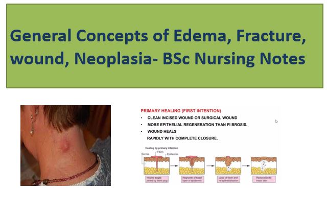
- Primary healing (healing by 1st intention )
- Secondary healing (healing by 2nd intention )
- Tertiary healing (healing by 1st and 2nd intention)
Primary intention
Secondary healing
- Heal with granulation tissue,
- These wounds take longer to heal resulting in
- form scar and
- re-epithelialization occurs primarily from wound edges
- Example
- pressure ulcers, dehisced surgical wounds
Tertiary intention
- When a wound is intentionally kept open to allow edema or infection to resolve or to permit removal of exudate, the wound heals by tertiary intention or delayed primary intention
- These wounds result in more scarring than wounds that heal by primary intentions
- but less than wounds that heal by secondary intention
Process of wound healing
- Hemostasis
- Inflammation Phase (0-3 days)
- Proliferative phase (2-24 days)
- Remodeling phase (24 days – 1 year)
- Process of wound healing
Factors influencing the healing process
Complications of wound healing
Neoplasia
- This term means new growth
- The new growth produced is called ‘Neoplasm’ or ‘Tumor’
- Not all new growth is neoplasm depends upon regeneration, repair, hyperplasia, hormonal stimulation, etc
- It is an abnormal mass of tissue, the growth which is excess and is uncoordinated with normal tissues
- “An abnormal mass of tissue formed as a result of the abnormal, excessive, uncoordinated, autonomous and purposeless proliferation of cells even after cessation of stimulus for growth which caused it”
- Oncology- Branch of science dealing with the study of neoplasms or tumors
- Neoplasm or Tumor is of two types
- benign
- malignant
Benign Tumors
- Encapsulated or well-circumscribed, small in size usually resembles the parent tissue
- Slow-growing and localised
- Noninvasive and no sign of metastasis
- Example- adenoma, papilloma, lipoma
Malignant Tumors
- Poorly differentiated
- Proliferate rapidly, spread throughout the body
- May eventually cause the death of the host
- Common term used for all malignant tumors is “Cancer”
- Example- Squamous cell carcinomas, Fibrosarcoma
TNM classifications
Staging of cancer is a system for describing the size and extent of spread of a malignant tumor, used to plan treatment and predict prognosis
- T- Tumor size
- N- Lymph node involvement
- M- Metastases
(Union Internationale Contre Cancer, Geneve)
T0 to T4–In situ lesion to largest and most extensive primary tumour
N0 to N3 – No nodal involvement to widespread lymph node involvement
M0 to M2– No metastasis to disseminated haematogenous metastases
Warning Signs of Cancer-CAUTION
C– Change in bowel or bladder habits
A– A sore that does not heal
U– Unusual bleeding or discharge
T– Thickening or lump in the breast or elsewhere
I– Indigestion or difficulty in swallowing
O– Obvious changes in the size of wart or mole or sore
N– Nagging cough or hoarseness