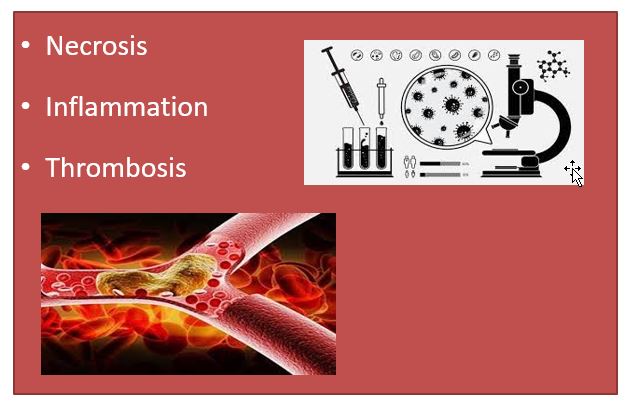
BSc Pathophysiology Nursing Notes of unit I on necrosis, Inflammation, Thrombosis
Necrosis
- Morphologic changes that follow cell death in a living tissue or organ
- Defined as a localized area of death of tissue followed by degradation of tissue by hydrolytic enzymes liberated by dead cells
- Necrotic cells cannot maintain membrane integrity and their contents often leak out.
- The enzymes responsible for the digestion of the cells are from lysosomes of dying cells or lysosomes of leukocytes as part of the inflammatory reaction
- Cell digestion by lytic enzymes
- Denaturation of protein in the cell
Morphologic alternation during the necrosis
Morphologic alternation
- Cytoplasmic changes- causing increased eosinophilia
- Nuclear changes- pyknosis/karyolysis/karyorrhexis
- Fragmentation and dissolution
- Discontinuity-Breakdown of the plasma membrane and organellar membranes
- Myelin figures- dead cells replaced by large phospholipid masses
- Leakage- Leakage and enzymatic digestion of cellular contents
Cause of necrosis
- Hypoxia
- Chemical and physical agents
- Microbial agents
- Immunological injury
Types of Necrosis
- Coagulative Necrosis
- Liquefactive Necrosis
- Caseous Necrosis
- Fat Necrosis
- Fibrinoid Necrosis
Coagulative necrosis
- The most common type and Mostly from sudden cessation of blood flow
- Due to denaturation of protein
- Characteristic of infarcts in solid organs
- This form of necrosis is where component cells are dead but the basic tissue architecture is preserved
- Commonly occurs in the heart, kidneys, spleen
- Example- Sudden occlusion of blood flow in coronary arteries (myocardial infarction) which leads to necrosis
- Bacterial and chemical agents can also cause but less often
Liquefactive or colliquative Necrosis
- Occurs due to degradation of tissue by the action of hydrolytic enzymes
- Whatever the pathogenesis, completely digests the dead cells resulting in the transformation of tissue into a liquid viscous mass
Causes of Liquefactive necrosis
- ischemic injury and bacterial or fungal infections
- Example- infracted brain, abscess cavity in gums
Caseous Necrosis
- The term “caseous” means cheese-like
- Derived from the friable yellow-white appearance of the area of necrosis
- The tissue is cheesy white in appearance
- Example- Tuberculous infections
Fat Necrosis
- Refers to focal areas of fat destruction
- Presence of shadowy outlines of necrotic cells
- Seen in the pancreas, breast
- Example- acute pancreatitis necrosis, traumatic fat necrosis
Fibrinoid necrosis
- Characterized by deposition of fibrin-like material in necrosed areas
- Example- autoimmune diseases, vasculitis
Inflammation
- Derived from Latin “Inflammare”
- This means burn and swelling
- Defined as the local response of living tissues to an injury
- It is a body defense mechanism which eliminates or limits the spread of injurious agents in the body
Why Inflammation?
The purpose is to
- To dilute, localize, and destroy the injurious agent
- To limit tissue injury
- To restore the tissue to normal
Causes of Inflammation
- Physical agents
- such as heat, cold, radiation, trauma
- Chemical agents
- such as acids, alkalis, poisons
- Infective agents
- such as virus, bacteria, toxins
- Immunological agents
- such as antigen-antibody reactions
Types of Inflammation
- Acute Inflammation
- Chronic Inflammation
Acute inflammation
- It is of short duration represents the early body reaction and is usually followed by repair
- Occurs vascular and cellular events, Migration, and Pavements
vascular events- chemotaxis
cellular events
- During cellular events, it occurs by leukocytosis and phagocytosis.
- Neutrophils are the act of first line of body defense, followed by monocytes and macrophages.
- When blood flow slows down leukocytes are pushed to periphery (margination and pavementing)
- Pro-inflammatory cytokines released by macrophages and mast cells causes endothelial cells and leukocytes to express adhesion molecules (selectins and sialyl lewis x proteins). These slows leukocytes and tumble along endothelium (rolling) and adhere with endothelial cells
- Transmigration of adherent leukocytes through endothelial gaps to extravascular space (Diapedesis)
- Once the extravascular space, leukocytes migrate towards the site of infection (chemotaxis)
- It is induced by chemokines produced by mast cells, complement proteins such as leukotriene B4, platelet factor 4, etc
- Neutrophils remain site of infection for 6- 24hrs followed by monocytes (24-48 hrs)
- Then leukocytes recognize dead cells or microbes and kill them (phagocytosis)
Signs of inflammation
- Dolor
- Color
- Rubor
- Tumor
- Functio laesa
Signs of inflammation
5 cardinal signs
- Rubor- Redness caused by vasodilatation
- Tumor- Swelling due to extra-vascular accumulation of fluid
- Calor- Heat caused by increased blood flow
- Dolor- Pain due to high pressure by accumulation of fluid/chemical mediators
- Functio latest- Loss of function (swelling and pain)
Consequences of Acute Inflammation
- Resolution- return to normal following acute inflammation
- Healing by scarring- when tissue is destroyed, healing occurs by fibrosis
- Suppuration- collection of pus
- Chronic inflammation- when acute inflammation persists, it may progress to chronic inflammation
Chronic inflammation
- It is an inflammation of prolonged duration (weeks/months/years) in which active inflammation, tissue injury and healing proceed simultaneously
Causes of Chronic Inflammation
- Following acute inflammation e.g. osteomyelitis, pneumonia
- Recurrent attacks of acute inflammation e.g, recurrent UTI leads to pyelonephritis
Clinical features of Chronic inflammation
- Mononuclear cell infiltration- macrophages, lymphocytes
- Tissue destruction- induced by inflammatory cells
- Proliferative change- repair (angiogenesis and fibrosis)
Types of Chronic Inflammation
- Chronic nonspecific inflammation
- Chronic granulomatous inflammation
- Chronic nonspecific inflammation
- Characterized by nonspecific inflammatory cell infiltration
- For example, chronic osteomyelitis
Types of Chronic Inflammation
Chronic granulomatous inflammation
- Characterized by the formation of granuloma
- For example, TB, leprosy
Thrombosis
- Process of formation of the thrombus within blood vessels
- Platelet deposition on the vascular surface is the initial stage of thrombosis
- Thrombus- solid mass consisting of platelets (thrombocytes) and fibrin in which red and white cells are trapped
Pathogenesis of Thrombosis
Virchow’s triad
Endothelial Injury
- Causes- ulcerated plaque, vegetation, mural thrombus, smoking, infectious agents, trauma, etc
- This disrupts the pathway of coagulation
Alternation in normal blood flow-stasis-turbulence
- Stasis- slowing of the circulation and a major factor in the development of thrombi
- Turbulence- an unstable movement that contributes to arterial and cardiac thrombosis by causing endothelial injury
- Normal blood flow is laminar
Stasis and turbulence cause
- Disrupts laminar blood flow and brings platelets in contact with the endothelium
- Prevent dilution by activated clotting factors
- Retard the inflow of clotting factor inhibitors and permits thrombosis
- Promote endothelial cell activation resulting in thrombosis
Hypercoagulability of blood
- Any alteration in the coagulation pathway that predisposes to thrombosis
- Less contribution
- Divided into primary and secondary
Hypercoagulability of blood cont.
- Primary or genetic-inherited causes such as mutation in factor V, Antithrombin III deficiency
- Secondary or acquired- pathogenesis such as MI, cancer, prolonged bed rest, use of heparin
Thrombi
- Thrombi are attached to the underlying blood vessels or heart wall at the point of origin
- Thrombi vary in shape and size of origin, cause, and pathogenesis
Types of Thrombi
- Based on color and components- platelet thrombus, coagulation thrombus
- Based on site and mode of formation- occlusive thrombus
- Based on infection- septic
Effects of thrombi
- They obstructs arteries and veins
- Formation of emboli
- For example- coronary thrombosis causes MI
Embolism
- When the thrombus travels from its site of origin resulting in partial or complete occlusion of a vessel causing smaller passage
- 90 % of emboli arise from thrombi and are called thromboembolic
Embolus
- It is a detachable intravascular solid, liquid, or gaseous mass that is carried by the blood to a site distant from its point of origin
Types of Emboli
- Thromboemboli (solid)
- Malignant cell emboli (solid)
- Ruptured atherosclerotic plaques (solid)
- Amniotic fluid (liquid)
- Droplets of fat (liquid)
- Parasites (solid)
- Foreign bodies such as silk, bullets (solid)
- Air embolism (gas)
Thromboembolism
- Lodging of thrombus and emboli in pulmonary circulation or systematic circulation
- Site of Origin of Thromboembolism
Types of Thromboembolism
- Pulmonary
- Systemic
- Air
- Amniotic fluid
Pulmonary Thromboembolism
Thrombus and Emboli lodge in Pulmonary Circulation
Systemic Thromboembolism
- Due to emboli that travel through the arterial circulation
Site of origin
- Heart- 80 % arises
- Aorta-20%
- Unknown origin (10-15%)
Air embolism
- Air forms as Emboli
- About 100 ml of air required to obstruct vascular flow
- Air may enter the venous during
- during delivery or abortion, air is forced into a ruptured uterine vein
- During artificial pneumothorax, hemodialysis,
- accidental opening of large veins
Fat embolism
- Fat globules in the circulation
- Causes
- fracture of the shaft of long bone,
- trauma of fat tissue,
- burn of adipose tissue
Pathogenesis of Fat Embolism
- Mechanical obstruction- micro aggregates of fat cause occlusion of pulmonary or cerebral microvasculature
- Chemical injury- free fatty acids released from globules cause toxic injury to vascular endothelium
- Thrombocytopenia due to coating of fat globules with platelets
- Amniotic fluid embolism
- Occurs during complications of labor and the immediate postpartum period
- occurs due to the infusion of amniotic fluid into maternal circulation via a tear in the placental membrane and rupture of uterine veins
- Characterized by sudden severe dyspnea, cyanosis, hypertensive shock followed by seizure and coma
